
Joseph, a fellowship trained, board certified orthopedic surgeon. Joseph Spine is an advanced center for spine, scoliosis and minimally invasive surgery. CT scans produce excellent detail used to diagnose osteoarthritis and fractures. People with metallic implants may not be able to undergo an MRI because of the strong magnetic field used in the test.Ī CT scan is better than an MRI for imaging calcified tissues, like bones. MRIs may also be used in cases where the X-rays are contraindicated, such as with pregnant women. An MRI can be better at detecting abnormalities of the spinal cord, bulging discs, small disc herniation’s, pinched nerves and other soft tissue problems. MRI scans are better for imaging water-containing tissue.

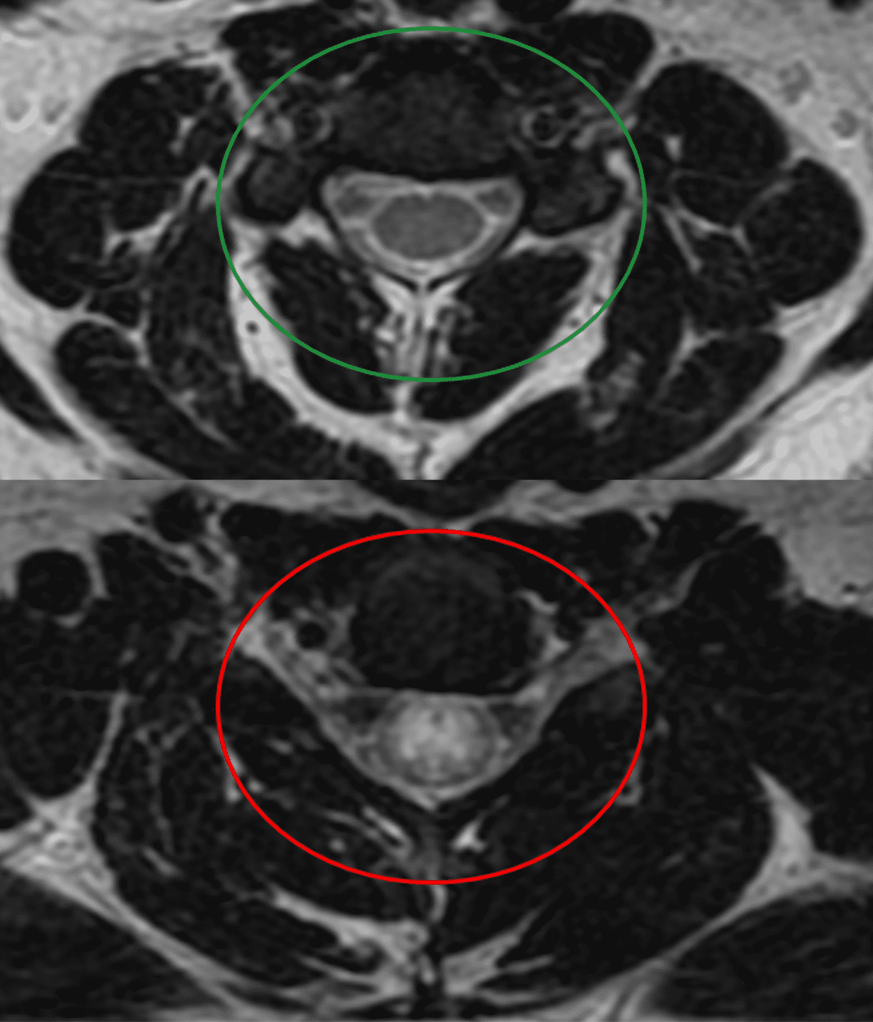
What the difference between an MRI and a CT SCAN?Īn MRI differs from a CAT scan (also called a CT scan or a computed axial tomography scan) because it does not use radiation. Imaging patients with metal (no magnet).Evaluating lung and chest issues (see lung scan image to the right).Pinpointing issues with bony structures (injuries).Imaging bone, soft tissue and blood vessels at the same time.The cross-sectional images generated during a CT scan can be reformatted in multiple planes, and can also generate three-dimensional images. Using CT, the bony structure of the spine vertebrae is clearly and accurately shown, as are intervertebral disks and, to some degree, the spinal cord soft tissues. CT images of internal organs, bones, soft tissue and blood vessels provide greater detail than traditional x-rays, particularly of soft tissues and blood vessels. Showing tissue difference between normal and abnormalĬomputed tomography, more commonly known as a CT or CAT scan, is a diagnostic medical test that, like traditional x-rays, produces multiple images or pictures of the inside of the body.ĬT scan is a rapid, 5-20 minute painless exam that combines the power of X-rays with computers to produce 360 degrees, cross-sectional views of your body.Imaging organs, soft tissue internal structures (see spine scan image to the right).It can give a very clear picture of the structure of the spine. The machine also makes a banging noise while it is working, which can make patients uncomfortable. Patients who are afraid this might happen should talk to the doctor beforehand, who may give them some medication to help them relax. There is a small theoretical risk to the fetus in the first 12 weeks of pregnancy, and therefore scans are not performed on pregnant women during this time.īecause patients have to lie inside a large cylinder while the scans are being made some people get claustrophobic during the test. Since radiation is not used, the procedure can be repeated without problems. The test is not painful you cannot feel it. There are no known dangers or side effects connected to an MRI scan. MRI also may show problems that cannot be seen with other imaging methods. In many cases, MRI gives different information about structures in the body that can be seen with computed tomography (CT) scan. Magnetic resonance imaging (MRI) is a test that uses a magnetic field and pulses of radio wave energy to make pictures of organs and structures inside the body. In actuality, both tests are valuable diagnostic tools that should be utilized by your spine care physician. They also wonder if one test is better than the other. Patients often inquire about the differences between an MRI scan and a CT scan.


 0 kommentar(er)
